The Celli Lab |
The Celli Lab |
|
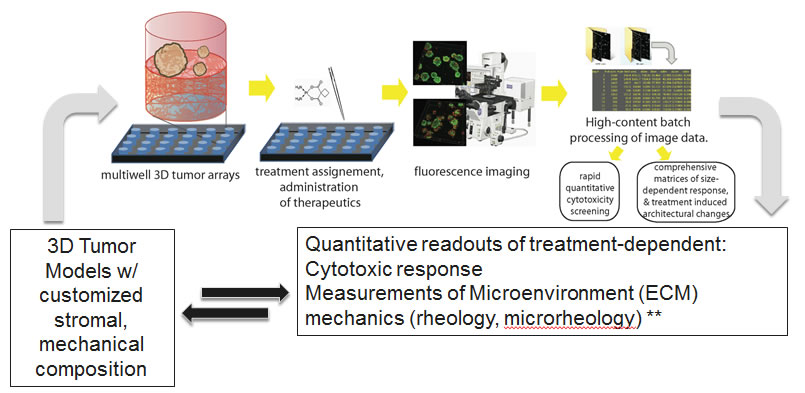
Cancer is a complex disease that presents myriad situations in which biochemical and biophysical interactions are inextricably linked. The growth behavior and therapeutic susceptibilities of cancer cells are influenced by the mechanical properties of surrounding tissue, which in turn remodel their physical environment in cooperation with stromal cells that play tumor-promoting roles. Motivated by this complexity our laboratory conducts translational cancer research using a multidisciplinary approach that combines physics-based analysis methodologies and therapeutic paradigms, especially photodynamic therapy (PDT), with traditional techniques from cancer biology. Measuring, monitoring and targeting mechanically sensitive growth behavior in 3D tumor models: towards rheology informed therapeutics 3D tumor models, in which cancer cells and stromal cells (e.g. fibroblasts) are embedded in customized formulations of extracellular matrix proteins allow us to examine the impact of matrix physical and biochemical composition on 3D growth behavior and response to interventions. At the same time we use in situ optical measurements of ECM mechanics (e.g. Jones et al, J Vis Exp 2014) to monitor the other side of this mechanoregulatory dialog in which cancer cells, in cooperation with stromal partners, remodel the mechanical microenvironment in response to external stimuli. This imaging-based approach to monitor changes in extracellular matrix mechanics is complementary and compatible with imaging-based therapeutic assessment in 3D tumor models (Celli et al, Scientific Reports 2014). This combination of methodologies allows us to explore mechanism-based therapies that target interactions with the mechanical microenvironment.
Targeting stromal crosstalk in pancreatic cancer with photodynamic therapy (PDT) PDT is a light-based treatment modality in which wavelength-specific activation of photosensitizing molecules selectively imparts cytotoxicity to tissue exposed to light. In PDT treatment of solid tumors, the PS is administered and allowed to accumulate in the malignant Low-cost enabling technology for PDT in global health settings A poignant example where PDT holds particular promise and applicability is the global health crisis presented by the high incidence of oral cancer in India. Although largely preventable, cancers of the oral cavity account for over 30% of cancers reported in India. This is one of the highest oral cancer rates in the world and is largely due to the widespread popularity of chewing gutka, a tobacco mixture with crushed betel nut and acacia extract. Treatment typically consists of surgery and/or radiotherapy, which require expertise and medical infrastructure that are often not available in the settings where they are most needed. Even if the disease is detected relatively early, these interventions can be disfiguring and present major quality of life issues including the ability to chew, swallow, speak, and work, thus increasing the societal economic burden on an already burdened economy. On the other hand, early clinical studies showed that PDT is a safe and effective approach, with remarkable healing and is especially effective for early stage cancerous and precancerous lesions of the oral cavity. Working with partners in the Hasan lab at MGH and Aligarh Muslim University, we are developing enabling technology for ALA-PpIX PDT and associated tumor imaging for lesions of the oral cavity. For more information see our recent publications on this topic (Hempstead et al, Sci Rep 2015; Mallidi et al J Biomed Opt 2015).
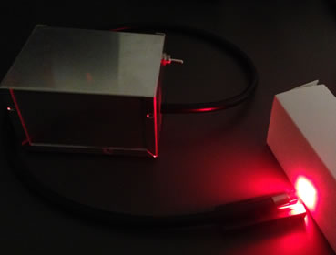
Above: a prototype battery-powered fiber coupled 635nm LED-based light source for PDT. | |